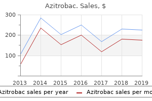
Discount azitrobac online mastercardSuccessful lateral retinacular release may be carried out using either arthroscopic or open strategies virus 0f2490 buy azitrobac amex. Preoperative Planning Range of movement antibiotics drinking buy generic azitrobac online, patellar tilt and subluxation antibiotics for staph discount azitrobac 100mg visa, and ligamentous stability must be examined whereas the patient is beneath anesthesthia antibiotics gram negative purchase 100mg azitrobac. The posteromedial and posterolateral compartments are visualized utilizing the Gillquist technique. Meniscal tears, articular cartilage lesions, and loose bodies are identified and addressed sugically when indicated. Once the diagnostic arthroscopy is completed, an Esmarch bandage is used to exsanguinate the leg, and the tourniquet is inflated. The camera is placed within the inferomedial portal and a hooked coagulation device within the inferolateral portal. If the superior lateral geniculate vessels are seen, they want to be aggressively coagulated. The surgeon should be succesful of tilt the patella 30 to 45 levels with the knee fully prolonged. After the discharge is accomplished, the tourniquet is progressively deflated to assess for excessive bleeding. The portal websites are closed, and a sterile compression dressing and cryotherapy system are utilized. The proximal starting point for lateral retinacular launch is just distal to the superolateral influx cannula. Arthroscopic view demonstrating profitable launch of the capsule and tight lateral retinaculum. Use of a cryotherapy system and a compression dressing will also lower the risk of hemarthrosis. The superolateral inflow cannula is a superb guide for essentially the most proximal place to begin of the discharge. Patients are initially seen 1 week after surgical procedure to assess knee movement and quadriceps operate and to take away sutures. The similar study found quadriceps energy deficits in 40% of sufferers, however in virtually all instances the energy was within 10% of the conventional leg. Patients might report a sensation of lateral instability if the patella sits in a medially subluxed place during early flexion, then snaps laterally throughout continued flexion. This is important because if the clinician incorrectly treats the presumed lateral instability with a medial stabilization procedure, the symptoms might worsen. Z-plasty lateral retinacular launch for the treamtent of patellar compression syndrome. Treatment of chondromalacia patellae by lateral retinacular launch of the patella. Lateral retinacular release: a survey of the International Patellofemoral Study Group. The patellar compression syndrome: surgical therapy by lateral retinacular release. Preoperative computed tomography scanning and arthroscopy in predicting consequence after lateral retinacular launch. If the knee stays flexed, the patella might remain dislocated over the lateral femoral condyle. The history of harm could additionally be unclear, especially if the patella rapidly and spontaneously lowered. In one cohort of 189 sufferers, 61% of first-time dislocations occurred during sports activity. On the opposite hand, patients presenting with recurrent patellar instability are much more likely to proceed experiencing further dislocations than sufferers who present with their first dislocation. The risk of a repeat dislocation in patients presenting with a historical past of prior patellar dislocation is about 50% over a 2- to 5-year period. Crosby and Insall3 reported that degenerative modifications had been uncommon after patellar dislocation.
Discount 500mg azitrobac free shippingAbout 30 to forty cc is injected intra-articularly treatment for dogs flaky skin order 250mg azitrobac visa, and about 5 cc is injected into every portal site antibiotic resistance mortality buy generic azitrobac 100mg line. The two most popular methods of leg assist are a knee holder (thigh immobilizer) and a lateral publish bacterial zoonoses purchase azitrobac amex. The knee holder should be positioned perpendicular to the position of the femur at a stage above the patella and portals that allows for a valgus force on the knee infection rate of ebola order azitrobac online pills. The end of the table is dropped down beneath 90 levels from horizontal to permit each legs to hold freely from the knees. The lateral post ought to be positioned above the patella and angled outwardly to enable for a valgus pressure on the operative knee. The surgeon ought to examine that the knee could also be taken through a variety of motion by abducting the leg towards the lateral submit with flexion of the knee off the facet of the desk. Preoperative Planning Before the surgery, all radiologic studies ought to be reviewed. The knee ought to be examined underneath anesthesia before starting the surgical procedure in an try to detect associated pathology. Padding of the contralateral leg is used to forestall pressurerelated injury to the bony prominences or superficial nerves. The superomedial portal is typically made proximal to the superior pole of the patella consistent with the medial border of the patella (medial to the quadriceps) and is directed in an oblique method into the joint. The anterolateral portal is created by making a small (about 6 mm) stab incision 1 cm proximal to the joint line and 1 cm lateral to the patella tendon. The anteromedial portal is considered the working portal for insertion of devices. It is typically made underneath direct visualization by inserting a spinal needle into the medial "soft spot" 1 cm medial to the patella tendon and 1 cm proximal to the joint line. Rasping may be carried out with either an arthroscopic shaver or a meniscal rasp that flippantly abrades both the tibial and femoral edges of the tear web site, as properly as the meniscosynovial junction, to stimulate vascularity. Trephination is carried out by inserting a long 18-gauge needle both percutaneously or by way of the arthroscopic portals across the meniscus tear to create vascular channels. The surgeon ought to keep away from perforation of the meniscus floor, causing additional damage. It is greatest used for posterior horn, center third, peripheral capsule, and bucket-handle tears. Before passage of the sutures, an incision is made posteromedial or posterolaterally to seize the needles as they exit via the capsule. For passage of a needle by way of the medial compartment, the knee is positioned in 20 to 30 levels of flexion to keep away from tethering the capsule. A 4- to 6-cm posteromedial incision is made just posterior to the medial collateral ligament, extending about one-third above and two-thirds beneath the joint line. Dissection is sustained anterior to the sartorius and semimembranosus musculature, deep to the medial head of the gastrocnemius. The posterolateral incision is made with the knee in 90 levels of flexion to enable the peroneal nerve, popliteus, and lateral inferior geniculate artery to fall posteriorly. A 4- to 6-cm incision is made simply posterior to the lateral collateral ligament, anterior to the biceps femoris tendon, extending one-third above and two-thirds under the joint line. Dissection is sustained between the iliotibial band and the biceps tendon after which proceeds deep and anterior to the lateral head of the gastrocnemius. On exposure of the capsule, a "spoon" or popliteal retractor is positioned in opposition to the capsule to visualize the exiting needles. A single- or double-lumen cannula is passed through the arthroscopic portals to the positioning of the tear. Long flexible needles are then passed by way of the cannula, piercing the meniscus above and below the tear site and creating vertical mattress sutures. Care is taken to not pull either suture throughout till both needles are handed. The sutures are then tensioned and tied to the capsule whereas viewing the repair arthroscopically. This method is greatest carried out on tears of the anterior and middle third, in addition to radial tears. The needle ought to enter the joint by way of the periphery to obtain a vertical or horizontal mattress suture configuration. A second needle with a wire retriever trocar is passed via the tear to retrieve the suture.
Order azitrobac cheapIn each circumstances the periosteum is incised bacteria under microscope cheap azitrobac line, and then a small cortical window is made with an osteotome yeast infection 9dpo purchase generic azitrobac on line. The cortical window is then replaced infection 68 buy discount azitrobac 500 mg line, and the periosteum is repaired over the defect antimicrobial wood sealer cheap azitrobac 500 mg without a prescription. The bone graft is impacted into the mattress, adopted by repeat discount and assessment of the chondral floor. Screw heads should be countersunk beneath the chondral surface to avoid hardware issues. For occasion, a compression screw may be positioned centrally in a lesion surrounded by absorbable pins on the periphery of the lesion to improve fixation. Final inspection should show a congruent discount with safe fixation of the lesion. Retrograde Drilling of the Femur Retrograde strategies are preferred as a result of they allow creation of a bigger channel for a more practical core decompression. Guidewire advanced to articular floor under fluoroscopic steering using retrograde method. As it approaches the articular floor, the drill bit should be advanced by hand for higher management. Two or three passes with the guidewire and cannulated drill bit are required for each lesion. Fluoroscopic picture of the cannulated drill bit over the guidewire utilizing the retrograde approach. Antegrade Drilling of the Femur Antegrade drilling of femoral lesions involves drilling from the articular surface into the lesion. Multiple drill holes are made immediately into the lesion utilizing a smooth, 1- to 2-mm guidewire to a depth that penetrates through the lesion and into healthy bone. Tibial lesions can be focused utilizing an anterior cruciate ligament information utilizing the retrograde approach. Guidewire is seen piercing the tibial articular surface using the retrograde approach. If unsure, a mini-arthrotomy should be made for direct entry and visualization of the lesion. Only lesions with involvement of subchondral bone with potential for subsequent collapse are clinically related. Daily range-of-motion workouts are encouraged, as a outcome of motion is important to present articular cartilage vitamin by way of synovial fluid diffusion. Patient compliance is a matter owing to the minimally invasive nature of the surgical procedure. A continuous passive motion machine could also be used for 2 to 3 weeks to help obtain movement. Physical therapy is concentrated on range of movement for the primary 2 weeks, after which light, progressive strengthening is initiated. Touch-down weight bearing is permitted through the first 6 weeks, adopted by progressive weight bearing. Radiographs are taken 1 to 2 weeks after surgery and on successive visits each 4 weeks thereafter. Once therapeutic is verified radiographically, the patient may be taken back to surgery for hardware removing if needed. The chondral surface could be inspected and the stability of the lesion can be evaluated at the moment. Most authors recommend elimination of any metal hardware on the joint floor to reduce secondary put on or attainable corrosion from synovial fluid. Therapy ought to give consideration to quadriceps strengthening and both energetic and passive vary of movement. Anderson et al used transchondral drilling to deal with 17 sufferers (20 knees) with open physes and 4 sufferers with closed physes. Skeletally immature sufferers had an 83% success rate, as opposed to 75% success in sufferers with closed physes. Failure to heal was associated with lesions in nonclassic locations, a quantity of lesions, and other underlying medical circumstances. Aglietti et al12 noted radiographic therapeutic in 16 knees, and all patients were asymptomatic at follow-up of 4 years.
500 mg azitrobac free shippingPatients with recurrent instability or episodes of giving means or those who are unable to return to activities of day by day living or sports activities are acceptable for surgical reconstruction bacteria 2 order azitrobac with mastercard. Patients with complaints of instability and a single-bundle or "partial" tear could profit from single-bundle augmentation antibiotic for mrsa order azitrobac 250mg visa, or double-bundle reconstruction in the occasion the remaining bundle is incompetent virus que crea accesos directos purchase 500mg azitrobac amex. Double-bundle reconstruction has been useful within the revision setting antibiotics drinking buy 100 mg azitrobac visa, significantly when the earlier femoral tunnel placement was within the conventional "over the top" place, which is too high within the femoral notch. This permits anatomic placement of the two femoral tunnels without interfering with the previous tunnel. Neither the peak of the affected person nor the scale of the knee has been an element when performing the surgery. Tibial tubercle Antromedial tibial incision Evaluation Under Anesthesia Range of movement in comparison to the contralateral knee Ligamentous examination Lachman Pivot shift Varus and valgus stress Anterior and posterior drawer Positioning the patient is positioned supine on the working desk, and the nonoperative leg is placed in a well-leg holder within the kidnapped lithotomy position. Injury patterns could embrace the following: Tear or stretch of one or each bundles Injury from femoral insertion, tibial insertion, and midsubstance There are 25 different injury patterns. The tibial footprints are left intact due to their proprioceptive and vascular contributions. Once the tip of the guidewire is placed in the right anatomic place (8 mm from anterior and 5 mm from distal articular cartilage), the knee is flexed to a hundred and twenty levels and the guidewire is manually tapped into the femur. The guidewire is over-drilled with a 7-mm acorn drill, taking care to avoid harm to the medial femoral condyle articular cartilage. On the tibial cortex, the tibial drill starts just anterior to the superficial medial collateral ligament fibers. If the location of the guidewire tip is unacceptable, the accent medial portal is used to insert the guidewire in the correct location. We favor to use two separate tibialis anterior or tibialis posterior tendon allografts. Alternatively, autogenous semitendinosus and gracilus grafts may be harvested (see Chap. The size of the EndoButton loop is chosen in accordance with the measured size of the femoral tunnels. The femoral fixation uses an EndoButton for every graft, and tibial fixation is obtained utilizing a bioabsorbable interference screw and Richards staple for each graft. Continuous passive motion is began immediately after surgery, from 0 to forty five levels of flexion, and is increased by 10 levels per day. From the first postoperative day, patients are allowed full weight bearing as tolerated. Non-cutting and non-twisting sports similar to swimming, biking, and working in a straight line are allowed at 12 weeks after surgery. Several short-term studies and a number of potential research currently are ongoing in Japan, France, Italy, and the United States. Rotational stability was achieved in each patient, as demonstrated by a unfavorable pivot shift. Preliminary outcomes have shown earlier return to full extension and symmetric flexion to the contralateral knee by three months after surgical procedure. Thus far, no vital tunnel enlargement has been found; nevertheless, follow-up has been short-term only. Two-bundle reconstruction of the anterior cruciate ligament utilizing semitendinosus tendon with endobuttons: operative technique and preliminary outcomes. Functional anatomy of the anterior cruciate ligament and a rationale for reconstruction. The effectiveness of reconstruction of the anterior cruciate ligament with hamstrings and patellar tendon: a cadaveric examine comparing anterior tibial and rotational loads. Anatomic reconstruction of the anteromedial and posterolateral bundles of the anterior cruciate ligament using hamstring tendon grafts. Reconstruction of the anterior cruciate ligament of the knee using a doubled tendon graft. Two failures have been sustained during contact injuries whereas taking part in collegiate football. The third occurred in a noncompliant patient 3 months after reconstruction when she returned to taking half in high school basketball without a brace. Bell et al2 performed biomechanical and pc modeling research comparing single and double femoral tunnels and the danger of femoral condyle fracture. Results of these studies have shown that fracture threat elevated significantly for the one tunnel versus the native condyle process, but no significant enhance in fracture danger was found for one versus two tunnels.
Cheap 250mg azitrobac with amexOsteochondral defects may cause decreased flexion via effusion antibiotic x 14547a cheap azitrobac 250 mg amex, or could have normal range of motion antibiotic for pink eye buy generic azitrobac. An isolated lesion may have point tenderness antibiotics used to treat lyme disease order azitrobac 500mg line, though it often is difficult to palpate antimicrobial breakpoints buy azitrobac now. Increased patellar mobility might indicate generalized ligamentous laxity, growing suspicion for patellar instabililty. Chondrocytes that produce the extracellular matrix are of mesenchymal stem cell origin. Chondral defects after a patellar dislocation may be found on the medial patellar side or lateral trochlea. Classically, osteochondritis dissecans occurs on the lateral aspect of the medial femoral condyle. Traumatic lesions could also be caused by compaction, as with an anterior cruciate ligament tear and lateral-based osteochondral damage, or by a shearing mechanism, as seen with patellar dislocations. Atraumatic lesions may be present in young persons, as is the case with osteochondritis dissecans, or in aged individuals, as seen with degenerative lesions. Traumatic, inflammatory, developmental, and ischemic causes have all been proposed but not confirmed. The pure history for juveniles with nondisplaced osteochondritis dissecans is very favorable. In one research, 81% of patients had tricompartmental gonarthrosis at a median of 33 years follow-up. Full assessment of the lesion was not completed until the defect was d�brided to secure rim. Large chondral defects will not be seen on plain radiographs, or could have a small radiodense bone fragment hooked up. Long-leg mechanical axis views are obligatory in sufferers with malalignment on physical examination, and should be thought of in all candidates for osteochondral autograft switch. Smaller lesions could also be amenable to microfracture or autograft cartilage transplant with single or mutliple plugs. Long-term studies could point out an elevated danger for degenerative arthritis with conservative administration,5 however no randomized managed studies exist. Nonoperative remedy ought to consist of physical remedy to obtain or keep painless, full vary of movement. Donors are screened with a multifactorial course of promoted by the American Association of Tissue Banks to reduce the chance of illness transmission. Preoperative Planning Mechanical alignment have to be assessed and, if essential, osteotomy planned for. Templated radiographs are obtained for applicable allograft sizing, based on the medial�lateral dimension of the lesion. Allografts are harvested within 24 hours of donor demise and can be preserved for up to 4 days at 4� C. Chondrocyte viability likely declines after 5 days, however prolonged storage-up to 21 days-currently is acceptable. Positioning We prefer to have the patient supine, preserving the foot of the desk up. A lateral publish and sliding footrest or taped sandbag permit for 90-degree flexion positioning of the knee. A tourniquet is positioned but is inflated provided that visualization is compromised by intra-articular bleeding. The defect sometimes is on the medial or lateral femoral condyle, requiring a longitudinal parapatellar tendon arthrotomy. Large trochlear or patellar defects amenable to osteochondral allograft transplation (rare) might require a larger parapatellar incision and eversion of the patella. A standard parapatellar arthrotomy is carried out to expose the defect on the affected facet of the knee. Sizing A the dimensions of the defect is decided using a cannulated cylindrical sizing device. Occasionally, a chondral defect is large or irregularly shaped, and requires multiple allograft.
Buy azitrobac 100mg overnight deliveryImportantly infection nosocomiale azitrobac 100mg low cost, particular attention have to be paid to gentle tissues such as plicae and the lateral retinaculum that doubtlessly may produce elevated compression between cartilage surfaces antibiotic 3 day course generic 250mg azitrobac visa. It is critical to d�bride all loose or marginally attached cartilage from the surrounding rim of the lesion virus in michigan generic 250mg azitrobac overnight delivery. A handheld tetracycline antibiotics for acne treatment buy azitrobac 500mg amex, curved curette is used to take away unstable and broken cartilage segments. A full-radius resector also may be used to remove unstable or broken cartilage from the lesion in preparation for the microfracture process. The calcified cartilage layer that continues to be as a cap to many lesions have to be removed, ideally through the use of a curette as famous by the blue arrow. This ready lesion has a stable perpendicular edge of wholesome, well-attached viable cartilage surrounding the defect, as famous by the green arrows. A properly ready lesion supplies a pool that helps hold the marrow clot-"super clot"-as it forms. A 90-degree awl is out there that ought to be used solely on the patella or other delicate bone. The holes are made as shut together as potential however not so close that one breaks into one other, thus damaging the subchondral plate between them. This technique often ends in microfracture holes which are roughly three to four mm aside. When fats droplets could be seen coming from the marrow cavity, the appropriate depth (approximately 2�4 mm) has been reached. Arthroscopic awls produce primarily no thermal necrosis of the bone in contrast with hand-driven or motorized drills. Microfracture holes are made around the periphery of the defect first, instantly adjacent to the wholesome steady cartilage rim (purple arrow). The microfracture holes are made starting on the periphery of the prepared lesion, maintaining the awl perpendicular to the bone. The microfracture course of is completed by making the microfracture holes (red arrows) toward the middle of the defect. The holes are as close together as attainable, three to 4 mm aside, however with none gap breaking into one other and disrupting the integrity of the subchondral bone plate. The quantity of marrow contents flowing into the joint is judged to be enough when marrow is noticed emanating from all microfracture holes. Finally, all devices are faraway from the knee and the joint is cleared of fluid. In these instances, and when the axial alignment and different indications for microfracture are met, first a few microfracture holes are made with the awls in various locations of the lesion to assess the thickness of the ebur- nated bone, and then a motorized burr is used to remove the sclerotic bone till punctate bleeding is seen. After the bleeding appears uniformly over the surface of the lesion, a microfracture process can be performed as described. We have observed noticeably improved outcomes for these sufferers with chronic chondral lesions since we started utilizing this technique. The black arrow points to a single microfracture gap that has been made to help assess the depth of eburnated or sclerotic bone that should be removed earlier than performing the microfracture process. Use a microfracture axe to make microfracture holes in the subchondral bone, first working all the best way across the periphery after which into the center of the lesion. Follow the rehabilitation protocol carefully to improve the chance of success. The machine often is ready at 1 cycle per minute, but the rate could be various primarily based on affected person preference and comfort. Crutch-assisted touchdown weight-bearing ambulation (10% of physique weight) is prescribed for 6 to eight weeks, relying on the dimensions of the lesion. Patients with lesions on the femoral condyles or tibial plateaus not often use a brace during the initial postoperative interval. This mobilization is essential in preventing patellar tendon adhesions and associated increases in patellofemoral joint reaction forces. Stationary biking with out resistance and a deep water exercise program are initiated at 1 to 2 weeks postoperatively.

Discount 100 mg azitrobac mastercardFull-thickness flaps are created from the midline of the clavicle both posteriorly and anteriorly antibiotic resistance purchase azitrobac on line, skeletonizing the clavicle infection 6 months after surgery azitrobac 500mg. After a distal clavicle resection antibiotic jock itch buy cheap azitrobac online, two 2-mm unicortical drill holes are placed within the posterosuperior surface of the distal clavicle buy antibiotics for uti online azitrobac 100mg otc, exiting through the intramedullary canal. One clamp is turned clockwise whereas holding the opposite finish till the sutures are intertwined collectively for the complete size of the sutures. The three units are intertwined counterclockwise in the same style, resulting in a twine of nine total sutures. If ligament augmentation was used, the ligament is wrapped in a determine 8 fashion and sutured to itself utilizing heavy nonabsorbable sutures. If the suture cord was used, the suture wire that was passed across the coracoid and thru the clavicle is tied. The ends of the suture twine are unraveled and each individual suture limb is tied to forestall unraveling of the cable. The sutures are positioned via the drill holes and tied excessive of the clavicle. If this is carried out correctly, the ligament should be taut and never overstuffed contained in the pocket. Draping is much like the open procedures, with extensive publicity and the arm draped free. An anterosuperior portal is made utilizing an outside-in method, utilizing a spinal needle to confirm positioning. D�bridement of the rotator interval is completed until the tip of the coracoid is visualized. Release of the superior glenohumeral ligament and partial release of the center glenohumeral ligament could also be required for adequate publicity. An anteroinferior portal is made near the tip of the coracoid, again with the outside-in method, using a spinal needle to confirm positioning and guaranteeing that the bottom of the coracoid could be reached using this portal. If the 30-degree arthroscope is used, the arthroscope place is changed to the anterosuperior portal. Drilling the assembled "Adapteur Drill Guide C-Ring with the Coracoid Drill Stop and Graduated Guide Pin Sleeve" (Arthrex, Inc. With the drill stop placed on the base of the coracoid (as near the scapula as possible), the corresponding area is marked on the superior aspect of the clavicle for the guide pin sleeve. Using the Adapteur Drill Guide C-Ring with the Coracoid Drill Stop underneath direct visualization, a information pin is positioned by way of the clavicle and coracoid, participating the drill stop. The pin must be centered on the clavicle and the coracoid and should exit the coracoid base as near the scapula as potential. The surgeon ought to take care to cease at the drill cease and not advance previous the coracoid base. The energy drill is indifferent and the cannulated drill is used as a portal to cross an 18-inch Nitinol suture passing wire. The limb of the Nitinol passing wire is introduced out of the anteroinferior portal, leaving the loop superior to the clavicle. The Nitinol suture passing wire is used to ship the white traction sutures via the clavicle and coracoid and out of the anteroinferior portal. While holding the blue TightRope suture tails, pulling on one of many white suture tails flips the oblong button to a vertical position, permitting passage of the TightRope via the clavicle and coracoid. Once past, impartial pulling on the white sutures flips the oblong button again to a horizontal position, anchoring it underneath the coracoid. Suture Passage and Tying the ability drill is indifferent, leaving the cannulated drill in place. After discount of the clavicle, sequential pulling on the blue TightRope suture tails delivers the round button all the means down to the superior clavicle, holding the reduction firmly. The blue TightRope suture tails of the spherical button are held firmly with one hand. Clavicle Reduction the surgeon pulls on the blue suture tails to advance the spherical button all the method down to the clavicle. An different is to displace the clavicle anteriorly with a towel clip to enable access for conoid tunnel drilling. Graft administration Semitendinosus ends are bulleted to allow for simple graft passage.
Buy azitrobac 250 mgOther muscles in and around the groin area additionally may be strained antibiotic resistance animals cheap azitrobac on line, including the rectus femoris infection 4 weeks after wisdom teeth removal purchase azitrobac 250 mg without a prescription, the sartorius herbal antibiotics for dogs cheap azitrobac 250mg fast delivery, and the stomach muscular tissues infection xbox generic azitrobac 500mg with amex, as can the conjoint tendon. However, with increasing experience, the examiner can really feel an irregular inguinal floor and appreciate abnormal tenderness contained in the external ring. In contrast, oblique and direct hernias involve easily palpable defects within the inguinal canal or through the anterior abdominal musculature, respectively. Duration of signs typically is months, and pain is proof against conservative measures. Osteitis pubis is characterised by symphysis ache and joint disruption and occurs commonly in distance runners and soccer gamers. It could additionally be troublesome to distinguish from adductor strains, and the two circumstances could coincide. Stress fractures are rare injuries that result from repetitive cyclic loading of the bone. Thorough information of the origins and insertions may be very useful throughout examination and palpation of the area. The posterior inguinal wall consists primarily of the transversalis fascia, together with the conjoint tendon, made up of the inner belly oblique and transversus abdominis aponeuroses. The pubic symphysis is a rigid, nonsynovial, amphiarthrodial joint consisting of layers of hyaline cartilage encasing a fibrocartilaginous disc. Eventually the medial thigh swells and ecchymosis is famous over the subsequent 2 to 3 days. Sports hernia is seen in aggressive athletes and occasional work accidents and will involve a selected traumatic episode, however most times is insidious and worsens over time with overuse. Coughing or Valsalva maneuver increases intra-abdominal stress and might increase tenderness, as can a resisted sit-up. The more than likely mechanism for osteitis pubis is that of elevated forces placed on the symphysis pubis from the pull of the pelvic musculature or repetitive stress from increased shearing forces. The ache is worse immediately during and after the exercise and improves with rest. These accidents normally happen along side an acute improve within the depth of training. The finger is inserted into the inguinal ring at the level of the exterior opening. The loose scrotal skin is invaginated and the finger is gently inserted into the exterior ring. The patient have to be requested for duration of symptoms, any inciting events, relieving and exacerbating elements, and timing of pain. To directly assess for hernia: In men: insert the finger into inguinal ring at stage of exterior opening. Gently really feel the inguinal floor and ask the patient to perform the Valsalva maneuver. Apply mild strain medially and laterally in search of abnormal asymmetric tenderness. In women: palpate the superior facet of the labia majora and upward to lateral to the pubic tubercle. Palpation of insertion of conjoint tendon: tenderness could improve, and a bulge could also be felt by having the affected person carry out a Valsalva maneuver. Hip flexion towards resistance: tests the energy of the iliopsoas and will detect a pressure or tear of this muscle. Herniography, which entails an intraperitoneal injection of contrast dye adopted by fluoroscopy or radiography, has been shown to identify sports hernias however has limited sensitivity and a substantial threat of perforation in up to 5% of sufferers. Once the patient can tolerate this, the major focus should be to regain power, flexibility, and endurance. Corticosteroid injection in osteitis pubis is controversial however could also be helpful in choose populations of athletic patients. Tissue repairs require longer rehabilitation and pose a larger threat for recurrence, primarily because of collegenases which are currently being described. Other mesh repairs fail because of surgical technique (eg, steel tackers, everlasting sutures within the periosteum, tight sutures involving nerves and causing necrotic tissue). The most rational and successful restore is using twolayered light-weight mesh, which supplies both posterior and anterior support and allows normalization of the torn anatomy.
|


